A 12 y o boy came with history of nocturnal seizures.
No neurological signs.
No neurological signs.
MRI BRAIN WITH MR SPECTROSCOPY


Findings:
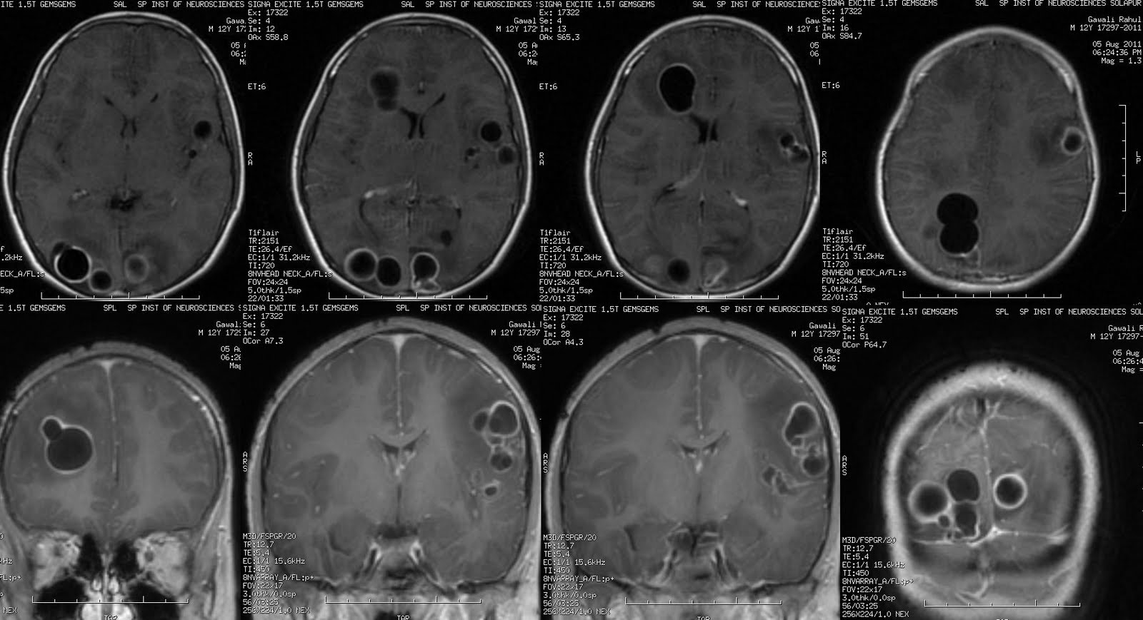
Multiple round to ovoid cystic signal intensity focal lesions in supra tentorium.
Content of cyst is very clear, iso intense to Csf on all pulse sequences.
No obvious eccentric nodule.
Most of the lesions show peri lesional vasogenic odema.
No significant mass effect. Effacement of hemispheric cortical sulci.
Enhancement is seen on post contrast T1w images along the thin T2w isointense wall with uniform thickness, most of the lesion show multi locularity. Size of cysts varies from 7mm to 2.5cm.
Single voxel MR Spectroscopy at short TE of 35ms and TR of 1500ms.
From right to left.
At 1.3 ppm - sharp short doublets of lactate.
At 2.01ppm - peak of NAA.
At 3.03ppm - no peak of Creatinine.
At 3.2ppm - no peak of Choline.
NAA/ Creatinine ratio is NA, Choline/ Creatinine ratio is NA.
Imaging wise possible DDs:
Abscess - Tubercular*, Pyogenic.
Hydatid cyst.
Cysticercoids unlikely as cyst size is too large with multi locularity. None of the lesion show eccentric scolex.
Hydatid cyst is possible though less likely as there is enhancement along its wall and perilesional odema, known in complicated or ruptured cysts and indicate ongoing inflammation. Usg abdomen for liver normal.
So Abscess, in that tubercular abscess is most likely.
No comments:
Post a Comment