Saturday 24 December 2022

AVN collapse prediction by Modified Kerboul method

MRI study of hip joints shows:
Avascular necrosis involving bilateral capital femoral epiphysis.


Modified Kerboul method is used for prediction of collapse in femoral head osteonecrosis by volumetric analysis on MRI. 

HOW IS IT CALCULATED ?

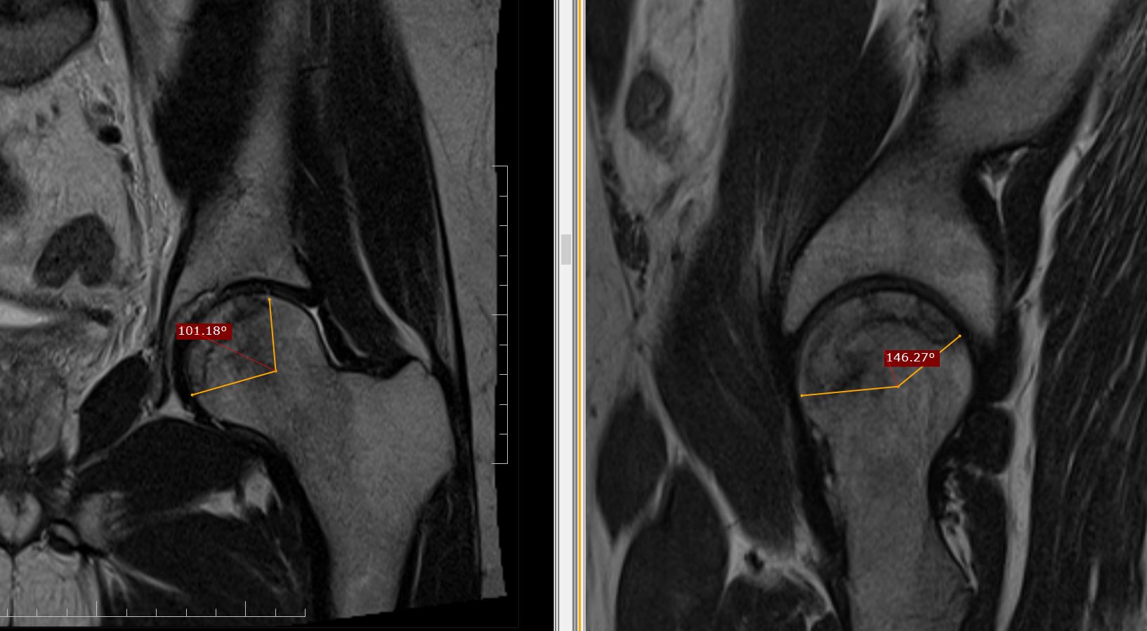
The arc of the femoral surface involved by necrosis measured by angles on midcoronal  (A) and midsagittal image (B) and then modified Kerboul angle (A+B) calculated by the sum of the two angles for both the joints.
Right side the angle is (129+169) =298, grade 3 lesion.
Left side the angle is (100+146) =246, grade 3 lesion. 

GRADING

On the basis of combined angle, hips are classified into four categories: 

Grade 1 (<200 degrees), 

Grade 2 (200 degrees to 249 degrees), 

Grade 3 (250 degrees to 299 degrees), and 

Grade 4 (>/=300 degrees).

BACKGROUND 

The hypothesis is that the combined necrotic angle measurement from magnetic resonance imaging scans predicts the subsequent risk of collapse in hips with femoral head necrosis.

With use of the modified method of Kerboul et al., Angle calculated by sum of the arc of the femoral surface involved by necrosis on a midcoronal as well as a midsagittal magnetic resonance image calculated on MRI, rather than on an anteroposterior and a lateral radiograph is far more accurate than on X-ray. 

Friday 4 November 2022

Physeal bony bar MRI

History of trauma 5 years ago. Operated with nailing for fracture of distal end of radius. 
Now presented with swelling and pain in the region of ulnar styloid process. 


MRI WRIST
  
Protocol:
 
Multi planner multi echo MRI study has been performed. Sequences planned are sagittal, Coronal and Axial FSE T1W images, sagittal, Coronal and Axial FSE T2W images, sagittal, Coronal STIR images.
 
This MRI study of wrist joint with x-ray correlation shows:
Clinical marker on skin.
Under growth of distal end of radius relative to ulna due to bony physeal bar, leading to positive ulnar variance of measuring approximately 15 mm, partial distal radio ulnar subluxation, leading to increased prominence of ulnar styloid process.
Abnormal abrupt angulation of flexor carpi ulnaris tendon over the prominent ulnar styloid process leading to changes of tendinosis owing to ongoing friction in the flexor carpi ulnaris tendon against prominent ulnar styloid process.
The bony physeal bar is involving midportion of growth plate of distal radius. Total width of growth plate measuring approximately 32 mm on coronal with bony physeal bar measuring approximately 11 mm in width in the region of linear track with low signal intensity foci of previous intra medullary nailing for distal end of radius. Physeal bar is involving nearly 30% of the total growth plate. Radial one third and ulnar one third of the growth plate intact.
There is 11° radial tilt on coronal and 23° dorsal tilt on sagittal of distal articular surface of radius.
V-shaped” groove involving distal articular surface of radius, proximal partial herniation of proximal carpal row in the distal radial groove.
Dorsal tilt of lunate bone measuring approximately 40° with dorsal shift of capitate axis.
No signs of lunate avascular necrosis.
 
Impression:
 
Bony physeal bar involving distal radius.

Tuesday 12 July 2022

Extramedullary focal fat - fluid level, a specific sign of osteomyelitis

 A 14 yo male with pain in calcaneum since 1months. 


MRI foot for calcaneum with CT correlation shows:

Heterogeneous signal abnormality diffusely involving calcaneum with multiple low signal intensity foci diffusely scattered in calcaneum on T1-weighted images which are hyperintense on STIR. Rest of the intervening calcaneum medulla shows faint high signal on STIR.

No obvious density abnormality on CT. No obvious sclerotic or lytic lesion. No obvious cortical destruction or sclerosis.

There is a focal lentiform shaped parosteal collection measuring approximately 26 mm in height and 6 mm in thickness medially at 2 o’clock position and 4 mm in thickness laterally on plantar aspect at 7 o’clock position on axial section. 

There is fat – fluid level within this collection, focal fat in the supernatant portion of this collection which is hyperintense on T1-weighted images with complete signal suppression on STIR, this portion follows classical fat density on CT. 

There is an associated diffuse oedema involving muscles of plantar aspect of foot especially quadratus plantae muscle, medial as well as lateral teno synovitis.

This finding suggestive of extramedullary focal fat - fluid level which is a pathognomonic sign of acute to subacute osteomyelitis.

Findings were discussed with the referring physician before finalizing the report, who added that there is elevation of inflammatory markers in lab reports and the suspicion of osteomyelitis clinically as well, with a feedback of significant improvement clinically after IV antibiotics. 

Take home note is during MSK MRI interpretation, a bone marrow signal abnormality with an associated focal periosteal extra medullary fat – fluid level, osteomyelitis should be in the list of your differential diagnosis.

References: 

1. Extra-osseous fat fluid level: a specific sign for osteomyelitis. Kumar J, Bandhu S, Kumar A, Alam S. Skeletal Radiol. 2007 Jun;36 Suppl 1:S101-4. doi: 10.1007/s00256-006-0194-1. Epub 2006 Sep 19.

2. Intramedullary and extramedullary fat globules on magnetic resonance imaging as a diagnostic sign for osteomyelitis. Davies AM, Hughes DE, Grimer RJ. Eur Radiol. 2005 Oct;15(10):2194-9. doi: 10.1007/s00330-005-2771-4. Epub 2005 Apr 29.