Showing posts with label Congenital. Show all posts

Friday, 15 May 2015

Syntelencephaly MRI

A 14 yo male child, birth history uneventful.Complaints multiple episodes of seizures since 10 months with history of developmental delay and clinical impression of cerebral palsy with epilepsy

On MRI, 
The interhemispheric fissure not visualized in fronto parietal region with midline fusion of the cerebral hemispheres, single lateral ventricle cavity. 
The sylvian fissure extending across the midline. Body of corpus callosum is deficient. 
Normal 3rd and 4th ventricle.
The fused superior frontal cortex is thick , has a agyric / polymicrogyric  pattern.

Diagnosis : Syntelencephaly.

A varient of semilobar holoprosencephaly _ Congenital malformation of brain also known as Middle Interhemisheric variant.
A rare malformation in which the cerebral hemisphere fails to divide in the posterior frontal and parietal region.

Similar case : Semi-lobar-holoprosencephaly

Contributed by Dr Mitusha Verma SR DNB Radiology Nanavati. 

Friday, 5 August 2011

'Wasp waist' sign of Congenital fusion on MRI

MRI Cervical spine mid sagittal T2 section shows a congenital fusion of C6-7 with rudimentary intervening intervertebral disc – ‘Wasp-waist’ sign.

‘Wasp-waist’ sign 

A radiographic appearance seen in congenital fusion of the vertebral bodies. 
Importance of this sign is to differentiate congenital from acquired fusion secondary to infection spondylodiscitis or surgery.
The antero posterior diameter at the level of the affected disco vertebral joint is smaller than the diameters at the superior and inferior limits of the vertebrae adjacent to the uninvolved discs.
Fused vertebrae form a continuous surface with concavity anteriorly. 

The term 'Wasp waist' refers to a woman's fashion silhouette, produced by a style of corset and girdle was very popular in 19th and 20th centuries. Its primary feature is the abrupt transition from a natural-width rib cage to an exceedingly small waist, with the hips curving out below. The sharply cinched waistline also exaggerates the hips and bust. To achieve these drastic measurements women suffered with cracked and deformed ribs, weakened abdominal muscles deformed and dislocated internal organs, and respiratory ailments. Displacement and disfigurement of the reproductive organs greatly increased the risk of miscarriage.

Tuesday, 2 August 2011

Arnold Chiari type I malformation MRI

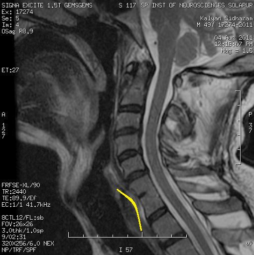
MRI Cervical spine sag T2 w image shows Pointed peg like low lying cerebellar tonsils by ~7mm. Obliteration of pre pontine cistern. 
No associated syrinx or signal abnormality in cord. 
No associated anomalies involving base of skull. 

Syn: Chiari type 1, Cerebellar tonsil Ectopia.

Definition: An abnormal caudal protrusion of the cerebellar tonsils across foramen magnum.
First described Hans von Chiari (1891).
There are total four types of Chiari malformations are described in the literature, type I, II, III, and IV.
Rest of the malformations are quite distinct imaging wise from type I. 

Imaging wise Diagnostic Criteria for Chiari type 1:
MRI, mid sagittal T2 section at the level of CV junction the best. 
1.  Low lying pointed and peg like cerebellar tonsils below the Opisthion - Basion line by more than 5mm for adult and 6mm for children. 
2.  The tonsillar descend should be primary and not secondary to or as a sequel of raised intracranial pressure by intra cranial space occupying lesion, hematoma or cerebral edema which is a seperate entity labelled as Acquired Chiari Malformation. 

Other associated finding on MRI: 
Pattern of sulci on cerebellar tonsills which are normally horizontal, gets vertical instead of horizontal. Effacement or obliteration of Cisterna magna and prepontine cistern.
There may be an associated focal Cord odema at CV junction or syrinx in cervico dorsal cord. Syrinx results from altered CSF dynamics, the exaggerated pulsatile systolic wave in the spinal subarachnoid space drives the CSF through anatomically continuous perivascular and interstitial spaces into the central canal of the spinal cord. Obstructive hydrocephalus is also known in severe cases.

Severity or degree of tonsillar ectopia measured and expressed in millimeters the tonsillar tip extends below the opisthion - basion line. Less than 5 mm does not exclude Chiari malformation, if other associated finding are present like syrinx, cervicomedullary kinking, elongation of the fourth ventricle, a pointed or peglike appearance of the tonsils present with patient clinically symptomatic. 
For children the descend or distance up to 3 mm is normal. For children this cut off criteria for diagnosis of Ch I is 6 mm.

Ch I is not directly associated with other congenital brain malformations.
However, bony CV junction anomalies are common in patients with Chiari I malformation like Platybasia, basilar invagination, Scoliosis , Retroflexed odontoid process, fusion, Atlantooccipital assimilation.
                               
DDS:
Acquired tonsillar herniation: Raised ICP secondary to intracranial space occupying lesion like tumour, hematoma, cerebral odema. Micro crania or Cranio synostosis. 
Pseudo-Chiari Malformation: an associated with sagging brain stem secondary to spontaneous intracranial hypotension and post lumbar puncture procedure. 

Monday, 1 August 2011

Temporal schizencephaly MRI

A 16 yo male for epilepsy evaluation. EEG report mentions left temporo parietal abnormal spikes.
MRI Brain:

Axial and coronal T2w images of Brain shows:
A cleft iso intense to Csf , extending from left temporal horn towards the caudal surface of left temporal lobe.
Imaging diagnosis : Left temporal schizencephaly, the site of cleft is unusual.

Schizencephaly

A congenital brain malformation characterized by cleft extending from pial surface of cerebral hemisphere to the adjacent ventricle.
Imaging wise best diagnostic clue is a gray matter lined cleft iso dense to Csf on CT and iso intense on all pulse sequences of MRI .
Location wise usually involves insula, adjoining pre or post central cortex. Temporal and occipital are atypical and very uncommon locations.

Types:
Type I (Closed lip) : Fused pial - ependymal seam lined by gray matter forms "furrow" in cortex.
Type II (Open lip) : Large or wide gray matter lined, fluid filled cleft.

Incidence: Unilateral more common than bilateral in live patient series where as bilateral clefts reported more commonly than unilateral in neuropathological series. Exact incidence not known.
Associated findings may be deformity of ventricle at the site of cleft, 'points' towards the cleft. Heterotrophia. Associated anomalies may be Pituitary or Optic pathway hypoplasias, Hippocampal malformation / mal orientation. Septum pellucidum may be absent, partially deficient or present. In temporal or occipital schizencephaly, an associated absence of septum pellucidum not expected.
MRI is preferred investigation of choice due to its multi planner capability and excellent grey white matter differentiation.

General pathological comments: Insult or inherited mutations lead to same pathologic / imaging features typically in MCA distribution  Intra uterine insult include infection, maternal trauma or toxin exposure. Reported with alloimmune thrombocytopenia.
Genetics: Mutation in homeobox gene Emx 2 in some (not all) cases, particularly type ii Schizencephaly. Emx 2 is the first identified mutation.

Clinical presentation: Patient with type I cleft may be near normal or may have seizures, motor deficit "congenital" hemi paresis.
Unilateral variety often present with seizures, or motor deficit "congenital" hemi paresis where as bilateral variety is associated with severe disability may present with hemi or quadraparesis, microcephaly, hydrocephalus, Spasticity, severe deveolpmental delay, mental retardation etc.
Age at the diagnosis depends on onset of symptoms like seizures or when the motor deficit / developmental delay is noticed by parents.

Reference : Diagnostic imaging Osborn.