Wednesday, 9 October 2024

Lateral hindfoot impingement MRI Ankle

MRI ANKLE

MRI coronal STIR section of ankle joint shows degenerative subchondral bone marrow oedema involving articular surface of lateral malleolus of fibula, lateral process of talus and calcaneum, loss of joint space, degenerative sub chondral cystic geodes in the lateral process of talus at subtalar joint without joint effusion.
The bone marrow edema in calcaneum is confined to the subjacent articulating surface and characteristically not present all along the cortex around the margins of the sinus tarsi on sagittal STIR. Possibility of lateral hindfoot impingement suggested.
If bone marrow edema in calcaneum is present all along the cortex around the margins of the sinus tarsi, this would not be characteristic and would instead suggest the different diagnosis, that of sinus tarsi syndrome.

There is associated diffuse thickening of anterior talo fibular ligament.

Osseous findings of lateral hindfoot impingement include degenerative changes like subchondral sclerosis, subcortical bone marrow edema at specific locations at the talus, calcaneus and or fibula, sub chondral cystic geodes in advanced cases.

The incidence of calcaneofibular impingement is somewhat lower than talocalcaneal impingement, with calcaneofibular impingement present almost exclusively in individuals shows changes of talocalcaneal impingement, suggesting that the talocalcaneal osseous changes occur before the calcaneofibular changes.


Monday, 17 June 2024

Dural ectasia spine MRI


Sagittal MR images of spine showing multiple contiguous widening of CSF space posterior to the cord in thoracolumbar region, associated posterior scalloping of the vertebral bodies, widening of caliber of the bony spinal canal, hypo plastic posterior elements. 

Axial T2 images, STIR coronal images showing multiple lateral meningoceles in thoracolumbar region with enlarged neural foramen.

Spinal cord displaced anteriorly, flattened, compressed against anterior confines of anterior bony spinal canal.


Imaging diagnosis: dural ectasia.


Dural ectasia is widening of the dural sac, associated with herniation of nerve root sleeves out of foramina. Scalloping of the posterior vertebral body, thinning of cortex of pedicles and laminae, widening of neural foramina expected to prolonged pressure effect from the dural sac containing CSF.

Dural ectasia is very well-known with  Marfan syndrome however can also be associated with other inherited connective tissue disorders, including Ehlers-Danlos syndrome and Loeys-Dietz syndrome, type 1 neurofibromatosis, ankylosing spondylitis, Lehman syndrome.

Dural ectasia can also be associated with trauma, scoliosis.

May be asymptomatic, may present with back pain, headaches, radicular pain, leg weakness or urinary incontinence.

Friday, 19 April 2024

Little finger FDP tendon, A2 pulley injury

 MRI LITTLE FINGER

SAGITTAL T2 5TH FINGER

SAGITTAL T2 5TH FINGER

AXIAL T2 A2 PULLEY AT MID PORTION OF PROXIMAL PHALYNX 5TH FINGER

This MRI study of litter finger shows: 

Full-thickness flexor digitorum profundus, FDP tendon tear, Zone II injury. 

There is associated retraction of the proximal end, spring coiled, the torn end of proximal stump is at the level of metacarpo phalangeal joint, with a gap of 36 mm. Length of the distal stump measuring approximately 20 mm from its insertion on to the base of distal phalanx. Quality of both torn ends of the tendon is good, sharp without abnormal fraying or degeneration.

Associated A2 pulley injury at the level of midportion of proximal phalanx with approximately 4 mm bowstringing of FDS. 

A3 pulley intact. 

No associated bony avulsion.

No associated joint subluxation.

No associated volar plate injury.

No associated collateral ligaments tear.

Sunday, 9 April 2023

Graefe Usher syndrome MRI


MRI brain shows:
Thinning of bony calvarium with inner table scalloping, cerebral cortical atrophy marked in bilateral frontal and temporal lobes with sub dural hygroma iso intense to Csf.
Diffuse cerebellar atrophy with widening of CSF space in posterior fossa overlying bilateral cerebral convexity, CP angle cisterns isointense to CSF.
Bilateral symmetric widening of bilateral internal auditory canal, Meckel's cave.
Widened empty sella.
Associated bilateral optic nerve atrophy, increased prominence of sub arachnoid spaces around bilateral optic nerves.

Imaging diagnosis: Graefe-Usher syndrome.

Usher syndrome is characterized by partial or total hearing loss, vision loss that worsens over time. The hearing loss is sensorineural, caused by abnormalities of the inner ear. 

A rare, congenital, autosomal recessive disorder characterized by retinitis pigmentosa and sensorineural hearing loss, first described by Von Graefe in 1858.

Saturday, 25 March 2023

Mazabraud's syndrome

A young patient presented with mild left thigh pain with swelling.


MRI with Xray correlation shows:

1. Mixed signal intensity lobulated lesion involving metadiaphysis of left proximal femur, part of adjacent epiphysis. Lesion is slightly expansile with groundglass matrix in the region of metaphysis on x-ray. No periosteal reaction on x-ray as well as MRI. No obvious pathological fracture. No abnormal adjacent bone marrow oedema on STIR.

Imaging wise possible diagnosis: Fibrous dysplasia.

2. Multiple T2 hyperintense lobulated space-occupying lesions involving muscles of left adductor compartment. The largest lesion measuring approximately 76 mm x 40 mm at a distance of 20 cm from greater trochanter on medial aspect of femur at 7 o’clock position on axial section.

Imaging wise possible diagnosis: intramuscular myxomas.

Intramuscular myxomas + left femoral fibrous dysplasia = Mazabraud's syndrome.

Saturday, 24 December 2022

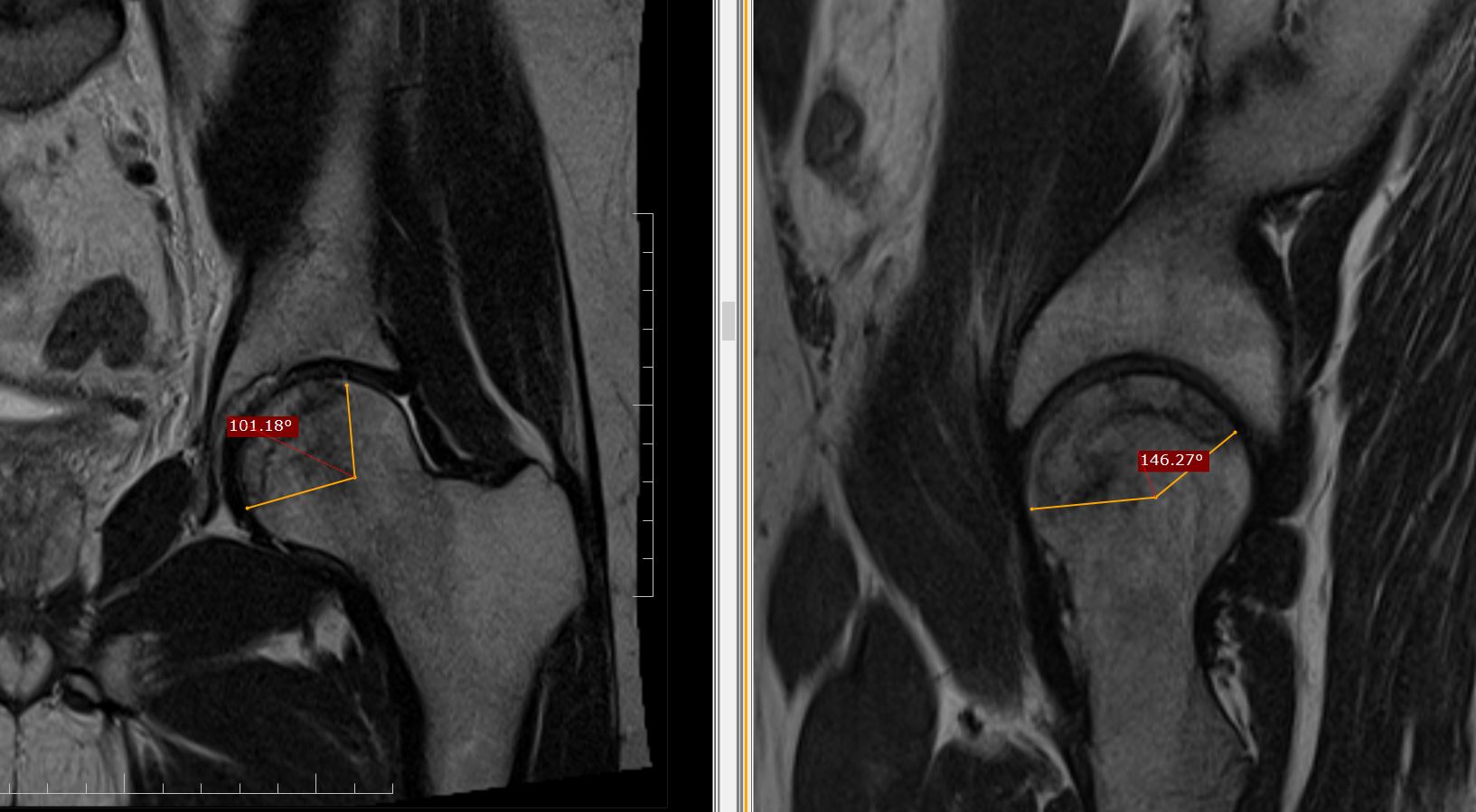
AVN collapse prediction by Modified Kerboul method

MRI study of hip joints shows:
Avascular necrosis involving bilateral capital femoral epiphysis.


Modified Kerboul method is used for prediction of collapse in femoral head osteonecrosis by volumetric analysis on MRI. 

HOW IS IT CALCULATED ?

The arc of the femoral surface involved by necrosis measured by angles on midcoronal  (A) and midsagittal image (B) and then modified Kerboul angle (A+B) calculated by the sum of the two angles for both the joints.
Right side the angle is (129+169) =298, grade 3 lesion.
Left side the angle is (100+146) =246, grade 3 lesion. 

GRADING

On the basis of combined angle, hips are classified into four categories: 

Grade 1 (<200 degrees), 

Grade 2 (200 degrees to 249 degrees), 

Grade 3 (250 degrees to 299 degrees), and 

Grade 4 (>/=300 degrees).

BACKGROUND 

The hypothesis is that the combined necrotic angle measurement from magnetic resonance imaging scans predicts the subsequent risk of collapse in hips with femoral head necrosis.

With use of the modified method of Kerboul et al., Angle calculated by sum of the arc of the femoral surface involved by necrosis on a midcoronal as well as a midsagittal magnetic resonance image calculated on MRI, rather than on an anteroposterior and a lateral radiograph is far more accurate than on X-ray. 

Friday, 4 November 2022

Physeal bony bar MRI

History of trauma 5 years ago. Operated with nailing for fracture of distal end of radius. 
Now presented with swelling and pain in the region of ulnar styloid process. 


MRI WRIST
  
Protocol:
 
Multi planner multi echo MRI study has been performed. Sequences planned are sagittal, Coronal and Axial FSE T1W images, sagittal, Coronal and Axial FSE T2W images, sagittal, Coronal STIR images.
 
This MRI study of wrist joint with x-ray correlation shows:
Clinical marker on skin.
Under growth of distal end of radius relative to ulna due to bony physeal bar, leading to positive ulnar variance of measuring approximately 15 mm, partial distal radio ulnar subluxation, leading to increased prominence of ulnar styloid process.
Abnormal abrupt angulation of flexor carpi ulnaris tendon over the prominent ulnar styloid process leading to changes of tendinosis owing to ongoing friction in the flexor carpi ulnaris tendon against prominent ulnar styloid process.
The bony physeal bar is involving midportion of growth plate of distal radius. Total width of growth plate measuring approximately 32 mm on coronal with bony physeal bar measuring approximately 11 mm in width in the region of linear track with low signal intensity foci of previous intra medullary nailing for distal end of radius. Physeal bar is involving nearly 30% of the total growth plate. Radial one third and ulnar one third of the growth plate intact.
There is 11° radial tilt on coronal and 23° dorsal tilt on sagittal of distal articular surface of radius.
V-shaped” groove involving distal articular surface of radius, proximal partial herniation of proximal carpal row in the distal radial groove.
Dorsal tilt of lunate bone measuring approximately 40° with dorsal shift of capitate axis.
No signs of lunate avascular necrosis.
 
Impression:
 
Bony physeal bar involving distal radius.

Tuesday, 12 July 2022

Extramedullary focal fat - fluid level, a specific sign of osteomyelitis

 A 14 yo male with pain in calcaneum since 1months. 


MRI foot for calcaneum with CT correlation shows:

Heterogeneous signal abnormality diffusely involving calcaneum with multiple low signal intensity foci diffusely scattered in calcaneum on T1-weighted images which are hyperintense on STIR. Rest of the intervening calcaneum medulla shows faint high signal on STIR.

No obvious density abnormality on CT. No obvious sclerotic or lytic lesion. No obvious cortical destruction or sclerosis.

There is a focal lentiform shaped parosteal collection measuring approximately 26 mm in height and 6 mm in thickness medially at 2 o’clock position and 4 mm in thickness laterally on plantar aspect at 7 o’clock position on axial section. 

There is fat – fluid level within this collection, focal fat in the supernatant portion of this collection which is hyperintense on T1-weighted images with complete signal suppression on STIR, this portion follows classical fat density on CT. 

There is an associated diffuse oedema involving muscles of plantar aspect of foot especially quadratus plantae muscle, medial as well as lateral teno synovitis.

This finding suggestive of extramedullary focal fat - fluid level which is a pathognomonic sign of acute to subacute osteomyelitis.

Findings were discussed with the referring physician before finalizing the report, who added that there is elevation of inflammatory markers in lab reports and the suspicion of osteomyelitis clinically as well, with a feedback of significant improvement clinically after IV antibiotics. 

Take home note is during MSK MRI interpretation, a bone marrow signal abnormality with an associated focal periosteal extra medullary fat – fluid level, osteomyelitis should be in the list of your differential diagnosis.

References: 

1. Extra-osseous fat fluid level: a specific sign for osteomyelitis. Kumar J, Bandhu S, Kumar A, Alam S. Skeletal Radiol. 2007 Jun;36 Suppl 1:S101-4. doi: 10.1007/s00256-006-0194-1. Epub 2006 Sep 19.

2. Intramedullary and extramedullary fat globules on magnetic resonance imaging as a diagnostic sign for osteomyelitis. Davies AM, Hughes DE, Grimer RJ. Eur Radiol. 2005 Oct;15(10):2194-9. doi: 10.1007/s00330-005-2771-4. Epub 2005 Apr 29.

Sunday, 17 October 2021

Rice bodies in subdeltoid bursa MRI


This MRI shoulder joint shows fluid distended sub deltoid bursa with numerous typical rice bodies.
Differentials given are rheumatoid arthritis and tuberculosis.
Suggested CBC, ESR, C-reactive protein, RA, anti-CCP in view of RA and joint fluid aspiration for culture and sensitivity in view of Tubercular Arthritis.

Rice body effusion or rice bodies in joint effusion or bursa represents an uncommon, nonspecific, inflammatory process, where multiple small loose intra-articular bodies resembling polished grains of white rice. 
They are typically seen with Rheumatoid arthritis, Tuberculosis. However it is also known to occur with juvenile arthritis, seronegative arthritis, osteoarthritis, septic joint, trauma and chronic bursitis.

They are almost of same size, hypointense on T1 as well as T2 weighted images without enhancement on post contrast, the shape is very typical with tapering ends resembling rice grains.
They should be differentiated from synovial chondromatosis where loose bodies are relatively larger in size, round to ovoid in shape and impart slightly high signal on T2-weighted images.

Elbow Neuropathic Arthropathy MRI

A middle aged male with unilateral left elbow pain, deformity, progressive swelling, restricted movement.

This MRI elbow joint shows:

Joint effusion, osteolysis involving proximal end of radius as well as ulna, articulating surface of capitulum as well as trochlea. Marked synovial thickening with frond like projections. 

Considering unilateral involvement possibility of infectious aetiology like Tubercular arthritis was one possibility would have been dealt with joint fluid aspiration for culture and sensitivity.
However, MRI CERVICAL SPINE screening was done with suspicion of neuropathic arthropathy which surprisingly revealed cervical cord syrinx.

Here is MRI CERVICAL SPINE of same patient which shows moderate cervical cord syrinx supporting neuropathic arthropathy.

So take home note is it is worthy to suspect and rule out conditions like this whenever applicable and possible as it considerably changes the line of management.

Friday, 8 October 2021

Steinstrasse CT KUB


This non-contrast CT study of abdomen for KUB of patient with left abdominal pain shows a dense large calculus in Urinary bladder which is in continuity with ribbon like column of multiple calculi in left ureter. 
Retrograde history taking unfurled that the patient had undergone ESWL one month ago.

Imaging diagnosis: Steinstrasse.

Steinstrasse is the German term which means "stone street", used to describe a possible complication of extracorporeal shock wave lithotripsy (ESWL) for urinary tract calculi, wherein a column of stone fragments forms that blocks the ureter.
Steinstrasse usually develops couple of months after ESWL and the most common site is the distal ureter.
The refined extracorporeal shock wave lithotripsy technique has reduced the incidence of steinstrasse from 20% to 6% 2.
Usually, the stone fragments pass spontaneously, but in about 25% of patients, retrograde stenting may be required. SOS more complex interventions such as stone flushing, ureteral dilatation, or long-term ureteral stenting may be required.

Sunday, 3 October 2021

Vitamin C deficiency mimicking inflammatory bone disease MRI hip

A 15 years old school goer presented with on and off bilateral hip pain, restricted hip joint movement, associated backache. 
MRI bilateral hip joint shows abnormal bone marrow edema involving bilateral acetabulum and subchondral portion of right sacral ala. Associated mild left hip joint effusion.
Imaging finding were suggestive of either inflammatory bone disease or multifocal osteitis. However the possibility of nutritional cause like Vit C or D deficiency was kept during discussion with the referring physician. Accordingly, getting the lab investigation done, was assured by refereeing doctor.

Patient's CBC, ESR, C-reactive protein was normal, RA factor was negative. Vitamin and mineral status showed normal zinc, folate and vitamin B6 levels. The vitamin C turned out strikingly low at 5 μmol/L (normal range 23–114 μmol/L) and vitamin D 25-OH at 12.2 ng/mL (normal > 30 ng/mL).
Treatment was already initiated by refereeing doctor with ascorbic acid, cholecalciferol, and liquid meal supplements keeping the diagnosis of scurvy on the basis of severely low vitamin C levels. 

The crucial message from this case is considering the nutritional cause for nonspecific bone marrow edema in MSK imaging is mandatory rather than confining our diagnosis between infective, inflammatory and neoplastic etiology all the time as in our case the treatable cause like Vitamin C deficiency was mimicking the inflammatory bone disease. 

Morel-Lavallée lesion MRI

Clinically RTA, run over by tractor.

This MRI study shows a focal well defined lentiform shaped subcutaneous collection on medial aspect of knee joint superficial to the superficial fascia. Collection is clear, hypo intense on T1-weighted images without any septations or loculations. No obvious high signal intensity methaemoglobin staining on T1-weighted images to suggest any frank haematoma.

Morel-Lavallée lesion

These are focal well defined lentiform shaped subcutaneous serous collections commonly encountered during MRI knee joint in the setting of severe trauma. However these lesions are typically described in thigh as a well defined collection overlying the greater trochanter of the femur and the tensor facia lata. 

These are actually localized haemolymphatic collections secondary to closed degloving injuries, where the skin and subcutaneous fatty tissue abruptly separate from the underlying fascia owing to trauma. The potential space thus created superficial to the superficial fascia is filled by serous fluid, some times frank blood. Similar collection secondary to similar biomechanical forces are described in lumbar region and over the scapula as well. 

The accumulated collection usually needs nothing to be done, resolves spontaneously. However may persist longer if it gets organized and encapsulated. The conservative management is with compression bandage. Surgical drainage may be sufficient for larger collection. However, the capsule may need to be resected to prevent recurrence if it is long standing and encapsulated collection with thick organized wall. 

Osteochondrosis of Superior Pole of Patella

Clinically young male patient with athletic background complaining of typical unilateral anterior knee pain. Marked tenderness at the superior pole of patella.


This MRI study of knee joint shows abnormal irregularity, fragmentation with sclerosis involving superior pole of patella. An associated thickening of quadriceps tendon. 
Knee joint effusion.
Imaging findings consistent with osteochondrosis of patella at superior pole.

Osteochondrosis of the superior pole of the patella

Osteochondroses are the heterogeneous group of injuries to the epiphyses and apophyses of children or adolescents, are actually osteonecrosis owing to repetitive microtrauma and avulsion injuries at tendinous insertions. Imaging wise characterized by bone fragmentation and sclerosis.

There are two well-known such syndromes associated with knee joint, one is Osgood-Schlatter disease, an avulsion of the tibial tuberosity and another is Sinding-Larsen-Johansson disease, a chronic avulsion injury involving lower pole of the patella at the insertion of patellar tendon.
The less well described osteochondrosis at the superior pole of patella appears secondary to similar mechanism associated with quadriceps tendon insertion, a rare cause of anterior knee pain in children between 5 and 10 years of age, usually single knee is affected but bilateral cases have also been reported.

Excessive lateral pressure syndrome MRI

This MRI Axial STIR sections of knee show clinical marker on skin on anteromedial aspect of knee joint. There is patellar tilt, articulating surface of patella facing medially, abnormal thickening of lateral patellar retinaculum and patellofemoral ligament. Associated bone marrow oedema involving lateral margin of lateral articulating facet of patella.

Imaging findings consistent with clinical diagnosis of Excessive lateral pressure syndrome.

Lateral Patellar Compression Syndrome

Synonym : Excessive lateral pressure syndrome, ELPS

This is another common cause of anterior knee pain.

The improper tracking of the patella in the trochlear groove generally caused by imbalance between medial and lateral dynamic stabilizers of knee, the tight lateral retinaculum restricting the patellar mobility with excessive lateral tilt of patella causing friction between lateral articulating facet of patella with lateral trochlea of femur.
Typically affects adults, patient presents with pain on compression of the patella, lateral facet tenderness. Condition is aggravated physical activity.

This is mainly a clinical diagnosis. However, lateral tilt of patella on axial sections of MRI or sunrise knee radiographs, patella facing medially without lateral translation should be depicted meticulously which is very commonly overlooked during MRI interpretation. Furthermore, abnormal thickening and shortening of lateral patellar retinaculum and lateral patellofemoral ligament could be appreciated on MRI. Nonetheless, the important ancillary findings on MRI are subchondral bone marrow oedema, cystic geodes involving lateral articulating facet of patella and adjacent lateral femoral trochlea facing the patella. The patellofemoral angle is calculated on axial sections, the medial opening of the angle, that is demonstration of angle more than 8° can support the diagnosis of ELPS.

Treatment is mainly conservative with physiotherapy focusing on quadriceps stretching and strengthening. Operative lateral retinaculum release is reserved for refractory cases. 

Discoid Meniscus


This MRI study of knee joint depicts discoid lateral meniscus.
No obvious associated meniscal tear or para meniscal cyst.

Discoid meniscus

This is a congenital condition and is bilateral in about 50% of the cases. Usually encountered as an incidental finding on MRI examination in about 5% of the cases, typically affecting lateral meniscus. Discoid medial meniscus is very rare.

The pathology behind  this discoid shape is loss of normal orientation of collagen fibres of meniscus.
Frequently this is an asymptomatic condition however the discoid meniscus has propensity for pressure, wear and tear leading early cystic degeneration, meniscal tear and para meniscal cyst formation. In such cases patient may present with knee pain with or without locking. 
MRI is the investigation of choice. 
The width of the body of meniscus, if 15 mm or more on coronal section is diagnostic of discoid meniscus. The body of the lateral meniscus is normally has bowtie configuration on sagittal section and seen only on two consecutive slices. If the meniscal body is seen on three or more consecutive slices while scrolling the sagittal sections, instead of normal bowtie tie should alarm of discoid morphology. 

The observation is usually managed conservatively if patient is not symptomatic. 
Partial or total resection is the option kept in front of patient if accompanied with complications like tear.

Supraspinatous Ganglion Cyst MRI

Clinically severe painful restricted shoulder movement, especially the abduction. Patient complaining that symptoms got aggravated over the period of time with shoulder exercise and physiotherapy. No obvious history of arthroscopy or intra articular injection.


This MRI shoulder joint shows a well-defined tear drop shaped cystic lesion along supraspinatus tendon tapering laterally towards its insertion suggestive of intra tendinous ganglion cyst. Associated changes of tendinosis involving supraspinatus tendon as there is mild tendon thickening. 
There was no sub acromial spur or para labral cyst.
Acromioclavicular joint normal.

The observation managed conservatively with intra-articular steroid. Clinical follow-up mentioned that patient improved clinically with range of movement improved by approximately 75%.
Suggested follow-up imaging

Imaging diagnosis: intra tendinous ganglion cyst of supraspinatus tendon causing shoulder impingement.

Trevor disease MRI

Clinical details, discharge summary mentions corrective osteotomy done for congenital progressive uni lateral knee deformity. Previous imaging details not available.
This MRI of knee joint shows post-operative status with corrective osteotomy for femur and tibia.
Well defined bone signal intensity outgrowth with cortex and medulla involving medial epiphysis of distal end of femur, epiphysis of tibial tuberosity with distinct cortex, medulla and fatty marrow in continuity with parent bone. Marked enlargement of patella with fragmentation.

Trevor’s disease

Synonym: Dysplasia epiphysealis hemimelica.

An extremely rare, approximately 1:1,000,000, congenital, non-hereditary condition consist of multiple osteochondromas arising from the epiphyses. Age of presentation is young children with slight male predilection.

There are three different types of involvement. The classic form has characteristic hemimelic distribution involving more than one bone or epiphysis within a single lower extremity. The localised form encompasses single bone involvement, either unilateral or bilateral. The generalised form incorporates the whole limb from pelvis to foot.
The abnormalities comprising the lower extremity is more common than upper, distal ends are more frequently encountered than upper ends of bone, medial aspects predilection is twice more common than lateral. The most common site of involvement is distal end of femur.

The condition typically demonstrates an irregular bone density mass on x-ray/CT arising from the epiphysis. The bony growth delineates areas of fatty marrow as well as foci sclerosis secondary to ossification on MRI. The illustration of continuity of cortex and medulla with the parent bone is must to end the search.

The bony growth can result in widening of joint space, deformity, bursa formation with soft tissue oedema, bursitis, tendinitis secondary to chronic irritation. The bony overgrowth can land up with fragmentation, detachment forming intra-articular bony loose body and changes of secondary osteoarthritis in advanced case.

Medial patellar plica syndrome

Clinically young patient presented with anteromedial knee pain.


MRI sagittal T2 and axial T2 images delineates a linear well-defined low signal intensity band running across medial patellofemoral recess. However, there is no obvious associated bone marrow oedema involving medial articulating facet of patella or medial femoral trochlea. Mild associated joint effusion.
Medial plica mentioned in the report with joint effusion.

Medial plica syndrome

Synonym: synovial plicae of the knee.

The another common cause of anterior knee pain typically present with pain on anteromedial aspect of the knee, just cranial to the joint line with or without associated with crepitation, catching and locking sensations. Typically involves young with athletic background. 

There are actually synovial invaginations as a part of remnants of embryological development. They are encountered in MRI over 70% of individuals and are mostly asymptomatic. However these tags can get inflamed secondary to repetitive friction and stretching, making patient symptomatic. They can undergo fibrosis after long standing inflammation imparting them non-stretchable restricting the joint movement and painful.
In symptomatic patients, medial plica seen as low signal intensity band on T1 as well as T2-weighted images with an associated chondral defect involving medial articulating facet of patella.
Treatment is mainly conservative, physiotherapy and intra articular steroid injections.

Sunday, 29 August 2021

Leukoencephalopathy, Calcifications and Cysts MRI


Clinically middle age male with history of progressive headache for last 5months.
CT brain shows multiple dense bilateral basal ganglia calcification. 
DDs thought were like Fahrs disease, hypo thyroidism, hypo, pseudo hypo para thyroidism.
However patients thyroid and para thyroid profile was normal and the posterior cranial fossa cyst could not be explained even by Fahrs. With a possibility of a concurrent posterior fossa tumor, patient was referred for MRI brain with contrast.
MRI brain with contrast showed dense calcification in bilateral basal ganglia. The posterior fossa cyst with debris level. No abnormal enhancement along wall of cyst or enhancing eccentric solid nodule depicted on post contrast MRI which ruled out tumor nodule. 
The bilateral symmetric confluent T2 white matter hyper intensity which represents an associated leukoencephalopathy.

Imaging wise primary diagnosis given was LCC. 

Due to mass effect, fourth ventricle compression by the posterior fossa cyst leading to obstructive hydrocephalus, patient underwent posterior fossa craniotomy and excision of posterior fossa cyst was done. Histopathology revealed nonspecific cyst without any tumor cells keeping with MRI diagnosis of LCC. 

LCC or  Labrune syndrome syndrome MRI

Leukoencephalopathy, brain calcifications, and cysts (LCC) also known as Labrune syndrome, an extremely rare with only near  10 cases reported so far in the medical literature.

The condition is caused by homozygous or compound heterozygous mutations in the SNORD118 gene on chromosome 17p13. Clinical presentation varies from spasticity, dystonia, seizures, and cognitive decline.

Etiopathogenesis of LCC is still a matter of debate. Obliterative microangiopathy has been found on histopathological examination as the basic abnormality, the cyst formation is due to necrotic process secondary to obliterative microangiopathy and calcifications is dystrophic in nature. White matter changes result from changes in water content rather than abnormality of myelination.

Another entity which deserves a special mention is cerebro retinal microangiopathy with calcifications and cysts is a distinct genetic disorder due to CTC1 gene problem. Similar leukoencephalopathy, cysts, and calcification have been reported in few cases in association with Coat's disease, described as “Coat's plus. Coat's disease is unilateral retinal telangiectasia with exudation commonly occurring in boys sporadically without systemic features. However, in Coat's plus, there is bilateral retinal telangiectasia with exudation along with systemic features in the form of LCC. 

In my case patient did not have any visual issues so that rules out retinal abnormality and the possibility of cerebro retinal microangiopathy.